Bad Things Ooze in Threes: Pyo Queen
- Nov 1, 2020
- 5 min read
Updated: Dec 3, 2021
You know what they say about how bad things come in threes? Well, we cut out third pyometra in three weeks.
The first was a young cat whose owner didn’t have the funds to spay her cat. She woke up to blood on her floor. The cat was diagnosed with an open pyometra, based on a combination of ultrasound, x-rays and blood work - and the bloody discharge coming from her vulva. Despite the electrolyte abnormalities, she was stable, therefore we waited over the weekend and her surgery went well on Monday morning.
In school, we were taught that the ovaries are the problem children. When a female ovulates, the follicle becomes a corpus luteum, sending a signal to the uterus to thicken the lining and prepare for a pregnancy. When a pregnancy does not occur, there is a small amount of pathology in the uterus that remains. Yet, dogs and cats do not have "periods" - in other words, they do not menstruate, and they do not shed the lining of the uterus as human females do. Dog owners mistakenly called the bloody discharge from the vulva of their intact female dog their "period" when that discharge is actually from the edema or swelling of the vaginal tissue, not from the uterus. If you remove the ovaries (via ovariectomy or ovariohysterectomy) then you do not get a pyometra.
As you can see from the ultrasound images, the uterine wall of this cat is very thick, and there is fluid inside the uterus that should not be there in a normal uterus. The only curative option for these patients is surgery.
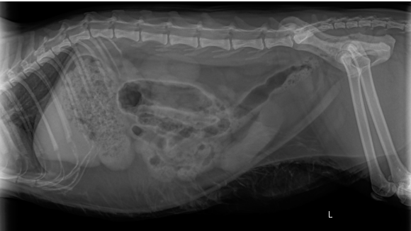
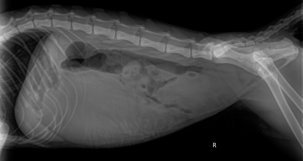
Not everyone will have an ultrasound machine however, so you may wish to take an x-ray. While it gives you an idea that there is an enlarged uterus, it is not always obvious. The radiographs here are the first case.
Can you see where the uterus lays?
How about these x-rays?
The second pyometra case was on the following Monday, late afternoon. An intact 8-year-old female cat who had stopped eating. I joked with our seasoned vet, saying that I was going to book him a pyo tomorrow - even before examining the pet. For the veterinary students who are reading, a pyometra should be high on the list of differentials for any intact female cat or dog who comes in not feeling well. For this patient, when I felt her abdomen during her physical, it was very taut with a focal mass in the cranial abdomen. I said to myself, ok, I need to ultrasound this, because I can’t convince myself this is uterus!

The above ultrasound image if a fluid filled structure. The wall of the organ is not thickened, however, this fluid filled structure is 5 cm in diameter - see the ticks on the y-axis. Wow! So what other structures in the abdomen are going to be this large and fluid filled - in the cranial abdomen??
Blood work showed a mild hyperglycemia, mild hyponatremia and mild hyperkalemia - likely due to shifting of the electrolytes into the third space. I have posted the CBC below for your learning opportunity. This shows a moderate to severe leukocytosis characterized by a neutrophilia and eosinophilia.

So, ultrasound, blood work and x-rays later, we scheduled her in for a spay the next morning.
Yes, that yellow stuff is pus --- inside the uterus. As I mentioned before, ALL non-breeding female dogs and cats should be spayed!!!
Our next case was a 6-year-old small breed female dog who presented as an acute abdomen case. She had been vomiting and then had weird breathing, which on exam was due to abdominal pain. The history was a bit vague, but I did get that she was not spayed. If you have forgotten, you can read my post on acute abdomen. So, we take a quick ultrasound and suspect she had a pyometra. This time the blood work was low normal glucose - which is borderline sepsis. She was not as bright as the other two patients. She appeared to be in distress. I was fortunate to negotiate a payment plan for this patient, as the family has limited funds, but the dog needed treatment - now. This was a case that could not wait. We started her with pain medication, IV fluids with dextrose and antibiotics, while our team scrambled to get surgical instruments and supplies ready. A note to your support staff - always be ready for an emergency surgery. A note to veterinarians working in a facility with no supplies ready - a disposable gown works as a make-shift surgical drape!
Since this patient couldn't wait a day, it was my turn to cut. Every surgeon says, a chance to cut is a chance to cure! Veterinarians know that a pyometra, especially a closed pyometra requires surgical correction. These patients will not survive without surgery. This is the case that really shows why... This dog had a septic abdomen. My RVT with years of emergency and critical care experience said it was the worst case of septic abdomen that she had ever seen. That definitely diminished my confidence in her survival. Also note, we do not have suction in our surgical suite. BUT, we gave her a chance as the owner was not willing to give up yet.
Here is what her ultrasound looked like:

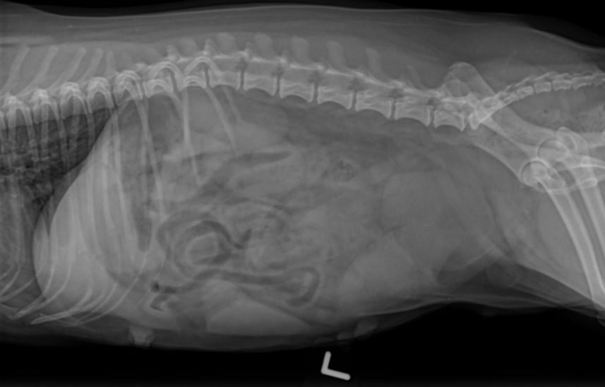
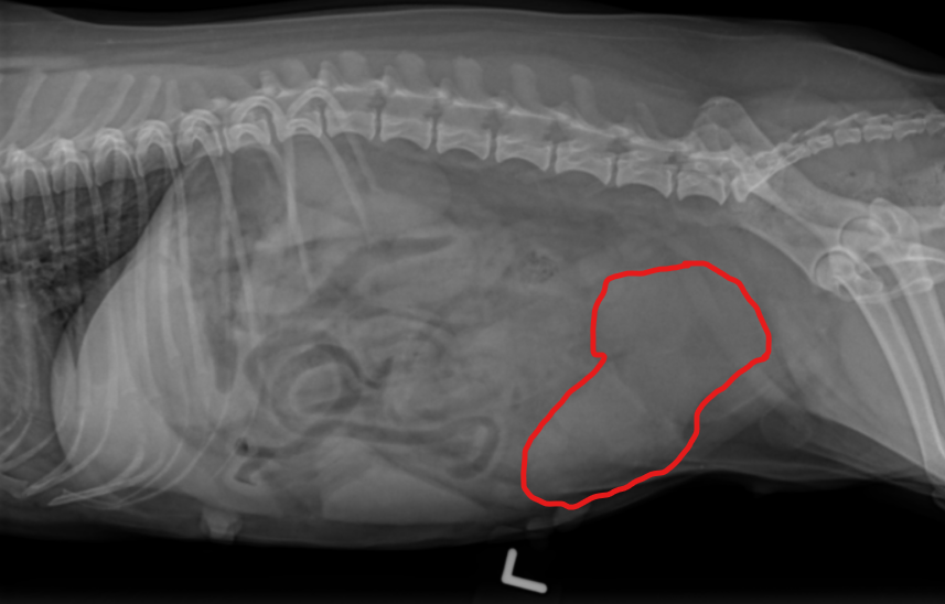
Again, if you do not have an ultrasound in your clinic, what the x-ray could look like:
So, we have a presumptive diagnosis of pyometra. Off to surgery we go!
After entering the abdomen, I can see that the uterus is already oozing. The material coming out was brownish red. We swabbed for culture and I collected for cytology while the RVT looked at that under the microscope, I was trying to exteriorize the uterus and some of this brown material shot out of the uterus across the room. *sigh*
Ok, our new graduate RVT is handling the anesthesia, doing a great job. I keep going. Telling her what I'm doing each step of the way. Ok, I'm going to be pulling on the ovarian suspensory ligament. We're still good. I manage to tie off both ovarian pedicles. Now, I'm looking at the tissue that is infected through to the serosal layer of the uterus. I decide that I want to tie off and remove the cervix. I look at my experienced RVT and I say, I want to take the cervix, I know if these were a rabbit that would be ok! What are your thoughts? She says, no one has asked me that! She calls someone at her ER clinic who says, yes, take the cervix and tie off as caudal as you can. Ok good! My thoughts were to take all the diseased tissue so that it wouldn't cause further problems. I tied off the uterine vessels individually, and then a circumferential ligature around the vaginal region. With the region packed off as much as I could, on excision, no disgusting brown material fell out. Phew!

OK, flush everything! My assistant brought warm saline for flushing. I flushed 10 times, blotted as much as I could. Anesthesia is still stable. She's not getting cold. Ok, we're closing up and going to hope for the best.
My assistant is cleaning up my surgical tray while I'm waiting for my patient to wake up. I mention that I swear I would have cleaned up my sharps if she hadn't gotten to it first! We have an on-going joke in the clinic as our most seasoned vet always leaves his sharps on the tray - so he owes them pizza every time he does (but hasn't paid up). So I say to the staff, if she survives to four days post-op I'm buying pizza.
We got pizza.
To read more on why your veterinarian recommends to spay all non-breeding females, click here.


Comments